Oooooh my favorites!
So the robust australopithecines have some interesting business going on. Their facial morphology is awesomely robust all with sagittal crests that served as attachments for the huge temporalis muscles, with what seems to be a pretty basic australopithecine both from the neck down. But here's the kicker, and something I honestly didn't know about three of the four robust guys we're dealing with here.
Their foramen magnum is heart shaped.
Yeah, my reaction too. Normally that feature is round, the point presents some interesting problems with the spinal cord and all of that but that would be pure speculation at this point.
To put it into perspective, here is a robust australopithecine next to a modern human skull.
And another with just the mandible.
To make it clear, the robusts are an evolutionary dead end, as in, they do not lead to modern humans. They did, however, live at the same time as early genus Homo.
Due to the stark contrast in the craniofacial morphology in the robust australopithecines, it's been proposed that they are different enough to deserve the genus name paranthropus. The jury is still out on that one for me.
Due to the bias in the fossil record, we have a bunch of skull and mandibular bits from these guys but very little post-cranial pieces. We do have the pelvis from Australopithecus robustus, and it's quite small, definitely not of the magnitude of bone thickness that the cranium has. This is also from the only robust australopithecine without a heart shaped foramen magnum.
It seems that Australopithecus aethiopicus has a sort of incipient heart shaped foramen magnum, it isn't full blown but it definitely isn't round. My thought is that this gave rise to Australopithecus garhi and Australopithecus boisei, due to the similar morphology and the similar geography. The question is whether or not Australopithecus robustus has the extreme facial morphology due to similar environment or similar ancestry. Au. robustus differs in its geography (it hails from South Africa rather and East Africa) and it's lack of the heart shaped foramen magnum.
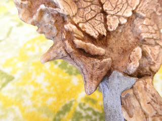
Here is the skull of Australopithecus boisei. You can clearly see VERY large zygomatics that flare out in order to accommodate a very large temporalis muscle. This muscle assists with bite force and must have been huge to need a sagittal crest to attach to in addition to the huge zygomatics. The environment was changing and the food was getting tougher so they needed to adapt. They accomplished this with increased bite force and tooth size.
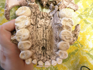
The OH5 dental arcade, maxillary, showing how large the teeth are. Those molars are about four times the size of our own and are completely worn down to the dentin from the gnashing action accomplished by a huge masseter and temporalis. A consequence of adjusting the bite force was the loss of prognathism. It drew the face in so it was more flat, and therefore able to apply more force to the tough foodstuffs they were eating.
Human Fossils and Evolution
Wednesday, March 9, 2011
Wednesday, March 2, 2011
Fun with Phylogenies
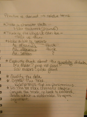
So I'm just going to toss photos of my notes from this week up here.
Biggest thing of note: when making a phylogeny for only one characteristic, that's kinda easy, it usually follows a time line type thing.
If only it were that easy.
In lab we ran into issues with the character state that we used (sagittal crest) as we ended up with a straight line. Once we went home to study group we came out with all kinds of possibilities on the white board when taking into account many different character states at once. We broke up into teams, made our own phylogenies, and then had to defend them to each other. Not such an easy task after all.
There's the how to.
You can see how we ended up with a time based line... not so insightful.
I feel like here we may have been caught up too much in the formalities of genus names and gave less consideration to actual characters, save for trying to stick Australopithecus sediba in there with some sort of logical place to go.
This one was backed up with many more morphological characteristics, and as a result ended up much more bush-like with many dead ends instead of a couple.
Wednesday, February 23, 2011
Australopithecus africanus
Australopithecus africanus (found in South Africa, where dating fossils is a wretched thing to behold,) has an interesting feature that I've become quite enamored with. They're called "anterior pillars." They appear to be some sort of reinforcement for some hard core chewing. They are unique to this species, and are just really cool. You can see them clearly on this specimen that we refer to as Mrs. Ples.
Look closely at the area right above the canines, there is a thickening that runs up the face. These are the anterior pillars.
So for some god-awful reason, our type specimen for Au. africanus, is a child, called Taung due to its discovery in a locality named Taung. That's pretty nasty, until you realize that the Taung child has a brain endocast, which allows us to see an imprint of the brain itself. Soft tissue preservation is rare due to the bias in the fossil record, and so this endocast is sort of special.
When you compare the Au. africanus pelvis to that of Au. afarensis, you'll notice that Au. afarensis has a noticeably longer pelvis. Au. africanus seems to be headed more toward the short and broad side of things here, which seems to show that the pelvis is becoming even more suited toward long term bipedal locomotion.
And if you take a look at the acetabulum, you'll notice that Au. africanus has a more complete and closed in acetabulum. It's still quite shallow, but is markedly more closed in than in Lucy's kind.
Another interesting feature on these guys is the fact that they begin to show some pretty large teeth. Large teeth become the focal point later on with the robust australopithecines and it seems like we're just getting closer and closer to crazy teeth. As the resources changed with the environment becoming cooler, drier, and more seasonal, obviously they needed to deal with the way they processed foods that were no longer squishy. Enter.... big teeth!
Thursday, February 17, 2011
Lucy (in the sky with diamonds?)
Lucy, also known as Australopithecus afarensis, is the rockstar of paleoanthropology. Ask anyone anything about the field and it seems Lucy is what they come up with. After all, she's super important, and insanely famous. She did something important for us, she showed the world that bipedalism and big brains did not evolve together, nor were big brains a requirement for bipedalism. This is HUGE, since we have for a long time assumed that this was one of those hand in hand sort of things. You can see the tiny braincase below.
So Lucy has a tiny brain and has a locomotor skeleton that is most definitely bipedal as evidenced by the bicondular angle present in the femur, as well as some of the characters present on the pelvis. The presence of the AIIS is another marker that helps support the case for afarensis' bipedal locomotion, since it is a unique attachment point of the illium that allows for muscles that aid in bipedal locomotion to attach.
The big thing to note here is the humongous amount of sexual dimorphism between the Au. afarensis individuals, as shown in the following photos.
Above are the femoral heads of two Au. afarensis individuals, there is a HUGE difference here.
On the left is a human femur, and it's pretty big. But check out the difference in size between the middle and right femur. Both Au. afarensis.
And there's the other end of that set up. Looks pretty much the same as a modern human, just much smaller.
One of the more compelling pieces of evidence for bipedalism in the Australopithecus afarensis species is the presence of the Laetoli Footprints. These are footprints made by this species in volcanic ash, that were then rapidly covered and preserved. This serves as a fossil of the soft tissue, allowing us to see much more than just the bones that we sometimes find.
There are thought to be three individuals represented by this fossil, two distinct tracks, with a third set of tracks right inside of one of the others.
Tuesday, February 15, 2011
Ardi
Have you met her?
Ardi (more formally known as Ardipithecus ramidus) is a pretty cool gal. She's got some weird stuff going on with her feet (more on that in a moment) and her pelvis looks like one of those 3D puzzles that take FOREVER to complete. She's also got some stuff going on with her teeth that isn't so common to find in the fossil record.
Ardi has this super cool foot. It's not totally like a human foot yet, but is totally capable of supporting bipedal walking, though in this weird tri-pod sort of way. The foot doesn't have a bony arch built into it, instead Ardi used her muscles to pull her foot up into an arch in order to be able to go for a stroll bipedally. There are a couple of bits of bone evidence showing this, where you can clearly see the grooves where the muscles ran through and would pull on to make an arch.
The cuboid bone here shows the groove beautifully.
This is the talus, which is the bone that sits in between those knobs on your leg that you tend to call your ankle. It looks a lot like a human talus, and yet, it belongs to Ardi.
This one is the cream of the crop. From left to right we see chimp, Ardi, and human feet. Ardi definitely has the ability to wrap the feet around as a chimp would and obviously has no bony arch like a human foot.
There is another important comparative feature that Ardipithecus ramidus has that I took photos of, and that is the form of the sciatic notch. In a chimp, we don't see this notch, but in a human and other hominin forms it's one of the things that we know causes our hips to be short and broad and accommodating to bipedalism. Ardi most definitely possesses this notch, and as Dr. Kramer pointed out, it may be the one single bit of really GOOD evidence for bipedalism on that insane puzzle of an illium that we have for Ardi because it hasn't been pieced together from tiny fragments and it is absolutely in the correct place.
This is Ardi's sciatic notch.
Here we have a human. If you compare the two you can actually see that though Ardi's is on a smaller scale, they actually follow about the same curvature.
And here's the chimp. Notice the lack of any real distinguishable sciatic notch, which allows for a much taller illium.
So there we have Ardi, a really interesting specimen that is still really new to those of us studying human evolution. It's a really cool stop along the path to life as we know it.
Tuesday, January 25, 2011
All About Orbits
(...with a little dentition for good measure...)
There are three major orbital enclosure that we are concerned about when dealing with primates. Two on the order itself, and one that helps us set them apart. There is a feature called a post-orbital bar that acts as a sort of enclosure to assist in stabilizing the eyeball within the skull of a prosimian, which is the primate most removed from us as humans. When you look at the skull of any monkey, ape, or human you would find that the eyeball is completely and securely enclosed within the skull, reducing eyeball movement even further. This helps increase visual acuity by reducing the amount of jostling around the eyeball does. It also helps with establishing full stereoscopic vision that is a feature we primates posses.
The photos below compare the orbital enclosure, clearly showing a post orbital bar in the case of the Galago (bush baby) skull, and the full enclosure of the eye in the Saimiri (squirrel monkey.) There is also a picture of a domestic house cat, showing the complete lack of any sort of post-orbital bar Instead the bone just sort of peters out around the eye.
Galago (bush baby)
Saimiri (squirrel monkey)
House cat (notice the bone not forming completely around the eye socket.)
Next I have a series of photos comparing the forms present in a modern day macaque versus Aegyptopithecus , which is an extinct form of monkey that was alive during the Miocene period. Note the amazing similarities between the two.
Front view of the two skulls. Macaque on the left, Aegyptopithecus on the right.
Notice the ever important size of the brain case. There has been some growth in brain size over the years.
Very similar dental pattern, as well as the placement of the foramen magnum, where the spinal cord exits the skull. Placement of this helps indicate just how upright some of these guys were.
As someone who works in a lab sorting fossils, I find a LOT of teeth. I'm talking probably 80% of the things I find and catalog are teeth. This definitely isn't a coincidence considering teeth are incredibly durable and made to outlast everything. They have to take a beating! They're gnashing up roots and tough things for goodness sake. They also have the fact that they are mostly inorganic material to begin with going for them, which means fossilization is faster since there is less organic material to replace. That said, you learn to both love and hate teeth. We've identified and named species before using only teeth that have been recovered from the fossil record. Without further ado, and lacking significant discussion because I'm new enough at this to still loathe teeth, here are some cool forms. My personal favorite of this bunch has to be the Notharctus dental impression, it looks incredibly modern. That's kind of a crazy thing to wrap your head around when you realize it's a prosimian from the Eocene.
Adapis teeth. Small Eocene prosimian with a sagittal crest.
Notharctus teeth.
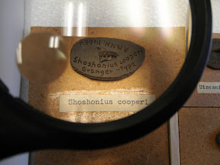
Shoshonius. Hard to tell from the teeth, but these Eocene prosimians had really huge eyes, which is one of our biggest indicators of having night vision capabilities. Prosimians are known for being nocturnal, which may have been a result of filling a vacant niche when monkeys started taking over the trees.
Until next week...
Thursday, January 13, 2011
Just a look-see
In lab on Wednesday we were able to take a look at the significant bones of the human body, and compare them to the same bones on monkeys and other apes. For me this was a bit of a review (thankfully!) and it was really nice to be able to take photos of the features that I find pretty interesting.
Here is a good front on shot of a gorilla skull. I wanted to be able to see the diastema, which is the spacing in the teeth created by the canines, which is there to allow the mouth to close properly.
Below are two photos that show the difference in the skull between a gorilla and human. Notice that the human skull lacks the sagittal crest, and that the sagittal crest on the gorilla is not purely a product of the left and right parietal bones meeting, but rather a feature that is present across both the frontal AND parietal bones.
Here I show the glenoid fossa, which is the part of the scapula where the humeral head fits into and rotates around in. Notice how shallow it is and the likelihood of shoulder dislocations. The shallow housing for the humeral head is a likely consequence of brachiation at some point in our ancestral past.
Next I have a comparison between the ankle region of a human and monkey. In the human we are looking at the left lateral and medial malleolus, the talus, and the calcaneous (heel bone.) The photo of the same region (though situated on the right side of the body) on the monkey shows that the calcaneous protrudes out much further than in the human.
Finally we have a human pelvis, and though the picture didn't turn out very well the feature that I was after turned out quite well. I want to draw attention to the sharp angle on the pelvis, that indicates this was a human male. The pelvic opening in a male is much less rounded than in a female due to the fact that a male doesn't need to push a baby head out through the opening. In a female the sacrum also isn't as sharply convex as in a male, also a consequence of child birth.
All in all this lab was a great step back into reminding myself where all of these wonderful bones and features are in the body. It's nice to be thinking about them in an evolutionary standpoint instead of a therapeutic one this time as well.
Here is a good front on shot of a gorilla skull. I wanted to be able to see the diastema, which is the spacing in the teeth created by the canines, which is there to allow the mouth to close properly.
Below are two photos that show the difference in the skull between a gorilla and human. Notice that the human skull lacks the sagittal crest, and that the sagittal crest on the gorilla is not purely a product of the left and right parietal bones meeting, but rather a feature that is present across both the frontal AND parietal bones.
Here I show the glenoid fossa, which is the part of the scapula where the humeral head fits into and rotates around in. Notice how shallow it is and the likelihood of shoulder dislocations. The shallow housing for the humeral head is a likely consequence of brachiation at some point in our ancestral past.
Next I have a comparison between the ankle region of a human and monkey. In the human we are looking at the left lateral and medial malleolus, the talus, and the calcaneous (heel bone.) The photo of the same region (though situated on the right side of the body) on the monkey shows that the calcaneous protrudes out much further than in the human.
Finally we have a human pelvis, and though the picture didn't turn out very well the feature that I was after turned out quite well. I want to draw attention to the sharp angle on the pelvis, that indicates this was a human male. The pelvic opening in a male is much less rounded than in a female due to the fact that a male doesn't need to push a baby head out through the opening. In a female the sacrum also isn't as sharply convex as in a male, also a consequence of child birth.
All in all this lab was a great step back into reminding myself where all of these wonderful bones and features are in the body. It's nice to be thinking about them in an evolutionary standpoint instead of a therapeutic one this time as well.
Subscribe to:
Posts (Atom)